What should be done when an examination detects breast abnormalities?
Take a deep breath. Don't panic! Learn about the results and treatment recommendations of mammography reports.
|
Category
|
Follow-up suggested treatments
|
|
0
|
Additional imaging may be required for assessment. Breast changes may occur
|
|
1
|
Negative: Regular examinations are recommended for 2 years.
|
|
2
|
Benign finding: Image changes are found but should be benign. Regular examinations are recommended every year. |
|
3
|
Probably benign finding: short-term (six months-a year) follow-up examinations are required.
|
|
4
|
Suspicious abnormality: Biopsy should be considered
|
|
5
|
Highly suggestive of malignancy: appropriate actions such as surgery or biopsy must be taken.
|
- The report results requiring an immediate return visit (referring to categories of 0, 4 and 5) are described as follows:
It is suggested that patients go to the division of breast surgery of the hospital immediately. Doctors will make appropriate treatment according to individual circumstances.
- Category 0: Additional imaging evaluation is needed.
As most Asian women have high breast density or there are other factors involved, it is not easy to distinguish abnormalities in mammogram and other imaging examinations are required. The common reexamination method is breast ultrasound, which is described as follows:
Breast ultrasound
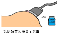
The method is to have the subject lie on her back with her hands raised high over the head. The doctor applies a hydrogel to the breast and scans clockwise or counterclockwise with an ultrasound probe pressed against the breast. Ultrasound is transmitted by a high-frequency probe. When it encounters tissues, it produces a reflection. The internal structure of the breast is transmitted back to the computer by the reflected echo. The image is integrated to show whether the lump is soft or a fluid-filled cyst. The non-invasive exam takes about 5 to 30 minutes, with no radiation.
Further breast ultrasonography reveals mostly normal or benign fibroadenomas. If any abnormalities are found, additional examinations are required to determine the lesion.

Schematic diagram of breast ultrasound examination
- Category 4: Suspicious abnormality – Biopsy should be considered
Suspicious might be malignancy Biopsy is recommended.
- Category 5: Highly suggestive of malignancy – Appropriate action should be taken
The findings have a high chance of being cancer. The most commonly established diagnostic method is interventional breast ultrasound and image-guided coarse needle biopsy (CNB), which is described in the following sections.
Coarse needle biopsy (CNB)
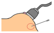
During examination, maintain posture and do not move. A local anesthetic is administered to the skin and four to six small pieces of breast tissue are cut with a thick needle (size 14 to 18) under the guidance of ultrasound or X-ray. It takes about 20 to 30 minutes. The wound, less than 0.2cm, must be pressurized and with an ice compress applied to for 1 to 2 days after surgery. There may be slight bruising, which will mostly subside in a few days. Risks and complications may include bleeding, infection, etc. To avoid delay in your treatment and diagnosis, it is recommended that you follow the physician's advice to receive necessary examinations.

Schematic diagram of CNB
Notes after examination:
- The wound should be compressed for 30 minutes. If there is blood stasis, warm towel can be applied 2 days later to relieve the symptoms.
- The wound should avoid contact with water as much as possible within one day after examination.
- Seek medical treatment immediately if there is inflammation (redness, swelling, heat, pain) or suppuration.
- The following are the results (categories 2 and 3) that require regular follow-ups without immediate return visits:
- Category 2: Benign finding
- Category 3: Probably benign finding
- Benign finding: such as calcification, fibroadenoma, fibrocystic breast, mastitis. What is calcification? Calcification in the breast is mainly calcium compounds in the breast tissue, which exist in the form of calcium salts. Calcification is not a disease in itself when it is found on mammography. Many benign conditions will be accompanied by calcification, such as vascular calcification, fibroadenoma calcification, or calcification caused by necrosis of breast fat cells after injury.
According to Health Promotion Administration statistics, the number of breast cancer cases in Taiwan is on the rise and has become the highest incidence of cancer among women in Taiwan. "Those who are not ill should not take it lightly, while those who are ill should not lose confidence."
Early detection and early treatment of breast diseases have achieved remarkable results. The most important thing is not to be an ostrich and refuse medical treatment, which might lead to further worsening condition! If you have any questions, discuss them with your breast cancer surgeon.

