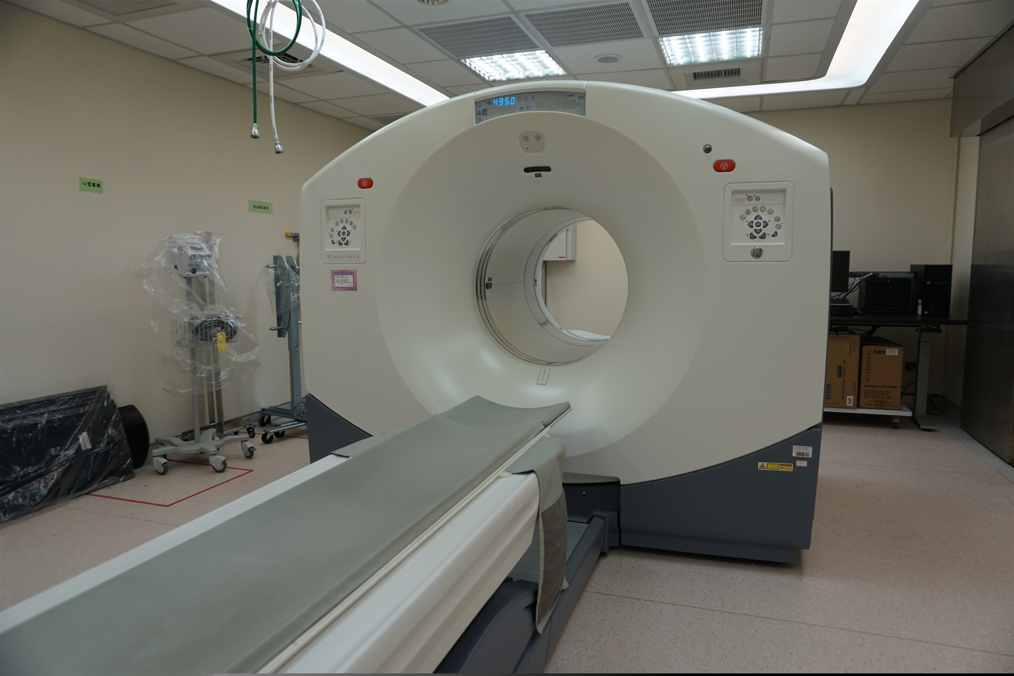
After the evaluation by your clinical physician, a positron emission tomography (PET) scan has been arranged for you. The following introduction is intended to help you gain further understanding of this examination.
The main purpose
- The application of PET/CT scans in health examination: a new modality for the early detection of cancer.
- The application of PET/CT scans in the field of oncology:
- Identification of malignant or benign tumors.
- Determination of cancer distant metastases.
- Evaluation of cancer treatment responses.
- Follow-up of cancer relapses or metastases.
Examination method
- The PET scan is a non-invasive functional nuclear medicine imaging modality that helps reveal how your tissues and organs are functioning. It applies a substance able to emit positive electron via its radioactive decay (i.e. the fluorodeoxyglucose, abbreviated as FDG, radiolabeled by F-18) to detect the glucose metabolism of cells in the human body.
- The tracer (i.e. FDG) is injected intravenously into your body. The tracer collects in areas of your body that have higher levels of metabolic activity, which often correspond to areas of disease (i.e. cancer cells). On a PET scan, these areas show up as bright spots. After computer restructuring, they form different contrast in images that can be read by naked eyes to make the correct diagnosis.
- In contrast to traditional examinations, PET offers a better detection rate and accuracy in a variety of cancers. The contemporary imaging systems are added with computer tomography, namely PET/CT, which further enhances accuracy.
National Health Insurance Indications
- Indications for Tumors:
- The staging, treatment, and suspected relapse or re-staging of breast cancer and lymphoma.
- The staging and suspected relapse or re-staging of colon cancer, rectal cancer, esophageal cancer, head and neck cancer (except brain tumor), primary lung cancer, melanoma, thyroid cancer, and cervical cancer.
- Staging: Evaluating tumor staging.
- Treatment: Evaluating the tumor response to treatment and planning to change the treatment strategy.
- Suspected relapse or re-staging: Applying to patients who have already received a certain phase of orthodox treatment and are detected with the level of relapse or metastasis and the evaluation of relapse (may not be used on routine follow-up examinations).
- The above steps must meet the following criteria: Those who could not be staged after conducting examinations such as CT, MRI, and nuclear medicine scan, or those who are recognized as limitations of CT and MRI to provide sufficient information for treatment needs, which must also be elaborated on the medical history of the indications for implementing positron imaging.
- Subjects who plan to cooperate with tumor treatment planning may use positron imaging as the treatment response evaluation tool. However, subjects who do not have subsequent and active treatment planning may not be arranged for a PET scan.
- Indications for Non-Tumors:
- Viable myocardium detection: Only applicable to those with a left ventricular ejection fraction of ≦ 40 % while the precise viable myocardium could (or recognized as) not be determined through traditional myocardial perfusion scan.
- Pre-operative evaluation of epilepsy syndromes: Pre-operative evaluation for those continuously and regularly take 3 or more types of anti-epileptic medications for ≧ 1 year and an average of more than 1 outbreak per month during the last 1 year with complications of loss of consciousness.
Non-National Health Insurance Indications
After injection, the radiopharmaceutical FDG is transported into a variety of inflammatory cells by upregulated glucose transporters. The utility of FDG for various inflammatory and infectious indications include:
- For patients with fever of unknown origin, it is used to detect underlying malignancies, infections or other inflammatory lesions.
- For patients with fever of unknown origin, it is used to detect underlying malignancies, infections or other inflammatory lesions.
- Diagnosing osteomyelitis, pulmonary inflammation/infection, orthopedic prosthetic infections, soft tissue inflammation/infection, or infections in other parts of the body, as well as monitoring the effectiveness of treatment.
- Discovering infectious foci in patients with bacteremia.
Precautions
- Pregnant women are advised not to undergo PET/CT scans. Please inform us before the injection if you are or might be pregnant.
- In the 6 hours preceding the FDG whole-body scan, avoid the ingestion or implantation of any substance containing calories, whether temporarily or permanently.
- Fast for 6 hours before the FDG whole-body scan (only plain water is allowed). Please inform if you are diabetic.
- During the 6 hours prior to the FDG whole-body scan, avoid administering any intravenous fluids containing calories (including TPN, PPN, Taita solution, Ringer's Lactate Solution, etc.). Only normal saline solution is permissible.
- Hemodialysis patients should refrain from peritoneal dialysis in the 6 hours preceding the FDG whole-body scan (due to the glucose content in the dialysate). Any glucose load (intravenous fluid such as TPN, PPN, Taita, Ringer's Lactate Solution, etc.; intraperitoneal dialysis) should be avoided 6 hours before performing a PET/CT to prevent competitive utilization with 18-FDG by target organs.
- If undergoing an FDG myocardial inflammation examination, please adhere to the following:
- 12 to 24 hours before the examination, consume two meals of a high-fat, high-protein, and carbohydrate-free diet (recommended items include boiled eggs and unsweetened pure butter).
- Fast for 12 hours prior to the whole-body scan (only plain water is allowed). Avoid intravenous fluids containing calories and peritoneal dialysis during the fasting period.
- FDG will decay over time so please inform the medical staff in case the subject needs to reschedule the examination time. Do not be late for registering at our department as it will affect the examination and time of the report.


